Ever wondered what the world looks like through the eyes of a shark? It's a realm of adaptation, survival, and sensory prowess far more complex than many realize. Dive in as we explore the fascinating world of shark vision, a sensory system honed by millions of years of evolution.
The sheer diversity of shark species from the colossal whale shark to the elusive Greenland shark means that their visual capabilities vary considerably. This variation is deeply connected to their specific habitats and hunting strategies. Some sharks patrol murky depths where light is scarce, while others thrive in sun-drenched coral reefs. The size and structure of their eyes are meticulously tailored to the demands of their environment, a testament to the power of natural selection.
| Characteristic | Description | Relevance to Shark Vision |
|---|---|---|
| Species Diversity | Over 500 known species of sharks. | Each species has adapted unique visual capabilities based on their environment and hunting style. |
| Habitat Variation | Sharks inhabit diverse aquatic environments, from shallow reefs to deep ocean trenches. | Eye size and structure are directly correlated with light availability in their habitat. Deep-sea sharks often have larger eyes to capture more light. |
| Eye Placement | Most sharks have laterally positioned eyes. | Provides a wide field of view, essential for detecting prey and predators from multiple directions. |
| Nictitating Membrane | Some species possess a protective eyelid-like structure. | Shields the eye from injury during feeding, particularly when hunting struggling prey. |
| Tapetum Lucidum | A reflective layer behind the retina. | Enhances vision in low-light conditions by reflecting light back through the photoreceptor cells. |
| Dermal Denticles on Eyes | Whale sharks have tiny tooth-like structures covering their eyeballs. | Provides mechanical protection and potentially reduces glare or biofouling. |
| Cornea and Lens | Work together to focus light onto the retina. | The cornea is a clear protective layer; the lens is a flexible structure for focusing. |
| Lens Shape | Unlike humans, sharks cannot change the shape of their lens. | Focus is achieved through movement of the entire lens, not through muscular adjustments of its shape. |
| Photoreceptor Cells | Rods and cones in the retina detect light. | Rod cells are more sensitive to low light, while cone cells detect color. Shark vision is generally considered monochromatic. |
| Eye Size | Varies depending on the sharks habitat and lifestyle. | Larger eyes are found in species living in darker environments, helping them to gather more light. |
| Visual Acuity | Sharks are capable of seeing very focused images. | Despite monochromatic vision, sharks possess surprisingly good visual acuity, enabling them to discern shapes and movements with precision. |
| Eye Parasites | Some sharks, like the Greenland shark, are prone to eye parasites. | The parasite, Ommatokoita elongata, targets the shark's eye as a habitat due to its protection from the outside elements. |
| Research Methods | CT scans and ultrasounds are used to study shark eyes. | These imaging techniques provide insights into the structure and function of shark eyes, helping scientists understand their unique adaptations. |
| Scientific Literature | Studies published in journals like PLOS ONE. | Peer-reviewed research provides valuable data on shark eye morphology, adaptations, and evolutionary significance. |
| Conservation | Understanding shark vision can contribute to conservation efforts. | Knowledge of their visual capabilities can help reduce bycatch and promote sustainable fishing practices. |
| Reference Website | Sharks.org | A leading resource for information on sharks, their biology, and conservation. |
Consider the whale shark, the ocean's gentle giant. Recent research, meticulously documented in a 2020 PLOS ONE report by Tomita et al., has revealed a remarkable adaptation in their eyes: they are covered in dermal denticles, tiny tooth-like structures. This novel mechanism of eye protection in vertebrates sets the whale shark apart. These eye denticles aren't just scaled-down versions of the dermal denticles found elsewhere on their bodies. They possess a distinct morphology, suggesting a specialized function, most likely offering mechanical protection against injury or perhaps even reducing glare. This discovery, made possible through CT scans and ultrasounds performed at the Okinawa Churaumi Aquarium in Japan, underscores the importance of ongoing research in unraveling the mysteries of shark sensory biology.
- Nsfw 18 Content What You Need To Know 2024 Guide
- Adam Garcia From Stage To Screen What You Need To Know
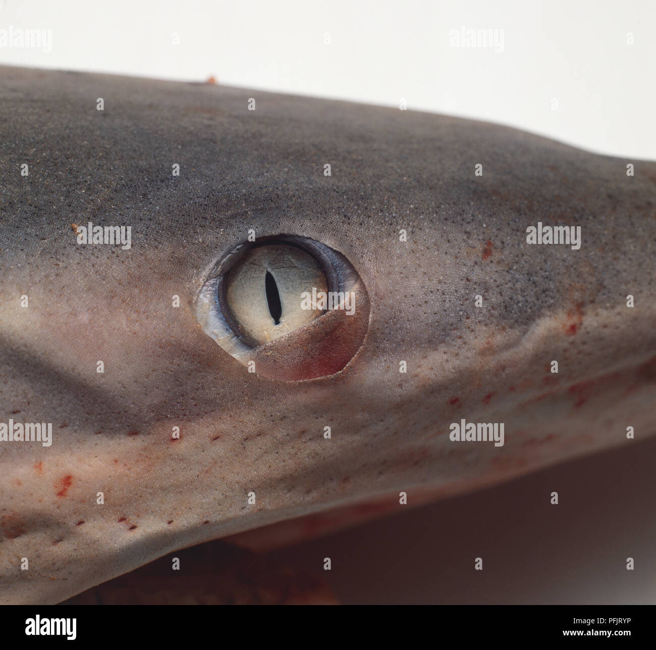
The eyes of sharks, like our own, consist of several key components: a cornea, iris, pupil, lens, and retina. The cornea, a clear protective layer, shields the eye from the external environment. Behind it, the lens focuses incoming light onto the retina, the light-sensitive tissue lining the back of the eye. However, unlike humans, sharks lack the ability to change the shape of their lens. Instead, they focus by moving the entire lens forward or backward, a mechanism more akin to how a camera focuses.
Many sharks, particularly those inhabiting murky waters or hunting at night, possess a tapetum lucidum, a reflective layer located behind the retina. This remarkable adaptation acts like a mirror, reflecting light back through the photoreceptor cells, effectively doubling the amount of light available for detection. This significantly enhances their vision in low-light conditions, granting them a distinct advantage over their prey.
While it was once believed that sharks were colorblind, recent research suggests that some species may possess a limited ability to perceive color. However, the majority of sharks are thought to have monochromatic vision, meaning they see the world in shades of gray. Despite this, their visual acuity, or sharpness of vision, is surprisingly good. They can discern shapes and movements with remarkable precision, allowing them to effectively track and capture prey.
- Who Are Storm Reids Parents Family Career Support
- Toni Kroos The Legend Retirement Footballing Achievements
The placement of a shark's eyes also plays a crucial role in its hunting success. Most sharks have laterally positioned eyes, meaning they are located on the sides of their head. This provides them with a wide field of view, allowing them to detect potential threats or prey from almost any direction. However, this lateral placement comes at the expense of binocular vision, which is the ability to see with both eyes simultaneously, providing depth perception. Some sharks compensate for this by briefly rotating their eyes forward to focus on a specific target, enhancing their depth perception just before striking.
One fascinating example of adaptation involves the nictitating membrane, a protective eyelid-like structure found in some shark species. This membrane can be drawn across the eye to shield it from injury, particularly during feeding. Imagine a shark attacking a struggling prey item; the nictitating membrane acts like a built-in shield, preventing the prey's thrashing from damaging the delicate surface of the eye.
The Greenland shark, a denizen of the Arctic's frigid depths, faces a unique challenge: parasitic copepods. These small crustaceans, known as Ommatokoita elongata, attach themselves to the shark's eyes, effectively blinding it. While this may seem like a significant disadvantage, the Greenland shark has adapted to this parasitic invasion. The parasite thrives in the shark's cold, deep environment, which lacks the complex fish immune systems that would normally evict such invaders. By specifically targeting shark eyes, Ommatokoita elongata has found a reliable niche, and the Greenland shark has learned to navigate its world despite its impaired vision, relying more heavily on its other senses, such as smell and electroreception.
The study of shark vision extends beyond simply understanding how they see. It has practical applications in conservation efforts. By understanding the visual sensitivities of different shark species, we can develop fishing gear that minimizes bycatch, the unintentional capture of non-target species. For example, research has shown that some sharks are attracted to certain colors, while others are repelled by specific wavelengths of light. This knowledge can be used to design fishing nets that are less likely to attract sharks, reducing the number of these magnificent creatures that are accidentally caught and killed.
The "gazing shark eyeball" mentioned in some contexts, particularly in gaming references like "Dave the Diver," highlights a different kind of fascination with shark eyes. In such games, these eyeballs might serve as upgrade components or crafting materials, showcasing the intriguing and sometimes morbid curiosity humans have about these creatures. While these fictional representations are far removed from the scientific reality of shark vision, they nonetheless reflect a deep-seated fascination with these apex predators.
The research on whale shark eye denticles, conducted at the Okinawa Churaumi Aquarium, involved a combination of advanced imaging techniques and careful anatomical analysis. Researchers used CT scanners to create detailed 3D renderings of the eye denticles, revealing their unique morphology and arrangement. They also employed ultrasound technology to examine the eyes of live whale sharks, providing insights into how these structures function in a living animal. This multi-faceted approach underscores the importance of interdisciplinary collaboration in advancing our understanding of shark sensory biology.
It's important to remember that not all sharks are created equal when it comes to vision. The size of a shark's eye, for example, is directly related to its habitat. Sharks that live in deep, dark waters tend to have larger eyes, which allows them to gather more light. Conversely, sharks that live in shallow, well-lit waters may have smaller eyes. The shape of the eye and the density of photoreceptor cells in the retina also vary depending on the species and its lifestyle.
Even the denticles on the rest of the whale shark's body, found on the head, trunk, and fins, differ from the eye denticles. The body denticles are characterized by parallel, triple ridges on their upper surfaces, while the eye denticles have a distinct morphology tailored to their specific protective function. This subtle but significant difference highlights the remarkable adaptability of these structures and their importance in ensuring the survival of the whale shark.
While a shark's eyeball might be similar to a human eyeball in its basic components cornea, iris, pupil, lens, and retina the details of its structure and function are finely tuned to the demands of its aquatic environment. The lateral placement of their eyes, the presence of a tapetum lucidum, and the occasional nictitating membrane all contribute to their unique visual capabilities. These adaptations allow them to thrive in a wide range of habitats and to effectively hunt and survive in the vast and often unforgiving ocean.
The study of shark vision is a complex and ongoing endeavor. As technology advances and new research methods are developed, we are constantly gaining new insights into the sensory world of these fascinating creatures. From the discovery of dermal denticles on whale shark eyes to the understanding of how Greenland sharks cope with parasitic infections, the field of shark sensory biology continues to reveal the remarkable adaptations that have allowed these apex predators to thrive for millions of years. The next time you think about a shark, remember that its vision is just one piece of a much larger puzzle, a puzzle that continues to intrigue and inspire scientists around the world.
- Bam Margeras Wild Ride From Jackass Star To Recovery Journey
- 2025 Indian Streaming Scene Mustwatch Hindi Web Series